Understanding the human mind is a millennia’s old pursuit, but it wasn’t until the 1970s that researchers were first able to peer into the brain without using a scalpel. Since then, researchers have been using powerful imaging technology - such as magnetic resonance imaging (MRI) and computerized tomography (CT) – to better understand cognitive diseases and disorders, help doctors better treat brain injuries, and predict recovery.
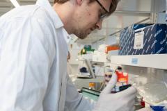
It's an area of research that captured Dr. Alexander Weber’s interest early in his career when he realized how versatile a tool MRI could be and how much he could discover about the inner workings of the brain using this technology. Unlike X-rays or CT scans, MRI doesn’t involve harmful radiation, making it safe to use for pregnant mothers, babies and patients that need multiple scans. Dr. Weber was particularly fascinated by the diversity of information an MRI can provide. He is studying how MRI techniques can be combined to tell clinicians something new about the brain and how that could lead to better treatments for children with brain injuries.
Earlier this summer, Dr. Weber joined the Brain, Behaviour and Development research theme at BC Children’s Hospital Research Institute as a new investigator. He is also an Assistant Professor in Pediatrics at the University of British Columbia.
We talked to him about his work and how imaging the brain is such an exciting frontier for research.
Why is MRI so important for medicine?
I think it still surprises people how much MRI scans have advanced over the years. They’re an incredibly versatile tool and we are still discovering how to improve them to better diagnose brain injuries or track whether treatment is working.
My research aims to improve and combine different ways of using MRI to give doctors the information they need to improve childhood health.
As an example, different methods of magnetic resonance imaging can highlight separate features of the brain, such as blood vessels or damaged tissue. Historically, MRI’s were only able to show contrasts in major tissue, allowing clinicians to roughly image the brain and tell if there were any large anomalies. Now, MRIs can show neural networks in much greater detail to give clues as to how the brain is functioning. They can even be used determine the presence and concentration of specific molecules in the brain.
How can new MRI techniques be used to help patients?
One way advances in MRI can help patients is demonstrated in previous research where I looked into concussions in ice hockey players. In this case, our research team was attempting to better image the fatty sheath that wraps around neurons, called myelin, which acts like the rubber insulation on an electrical wire.
We were measuring whether the specific signal from fluid between the myelin sheath and neuron was reduced after a concussion, as this could have significant implications for how biological signals move down the length of the nerve. We didn’t know if the myelin sheath was loosening or if it was degrading – such as what happens in Multiple Sclerosis. Using two different types of MRI imaging we found that this reduction in fluid signal was because it was loosening, which could have a big impact on how the injury is treated and what the prospects are for recovery.
Further research could enable a doctor to use these MRI techniques to better advise a hockey player whether they need to wait a few weeks before going back on the ice or if they need to stop playing for longer.
What is the aim of your current research?
My main focus now is brain injuries in newborn babies.
These injuries usually occur when there has been some sort of traumatic injury during birth such as a lack of oxygen, known as hypoxia, or brain swelling, known as encephalopathy.
When babies are born with a lack of oxygen it’s important that doctors have the information they need to understand the extent of the damage to the infant’s brain. Conventional MRIs that are usually routine in these cases struggle to distinguish between different types of brain injury, such as damage to white matter (the neurons in the brain) or brain hemorrhages. Our research aims to improve MRI scans so they can better tell the difference between these two.
Knowing whether abnormalities on a scan are due to white matter injuries or bursting blood vessels could dramatically change how clinicians treat the injury as well as the prospects for recovery.
Another method that we’re working on is known as Quantitative Susceptibility Mapping, or QSM analysis. This method allows us to measure how much oxygen is being used within the brain of an infant, which is a really good indicator for brain health. We can quantify how much oxygen is available in blood as it goes into the brain and subtract from this the amount of oxygen coming out. So this gives you an idea of how much the brain is using. An injured brain is likely to have a reduced metabolism so you would expect to see more oxygen left over. However, calibrating this equipment to measure oxygen does involve a slightly invasive procedure, so if we can work out how to make this less invasive it could be a far better tool to work with.
What got you interested in this field of research?
I’ve always been really interested in brains and how they work. The turning point in my academic career happened during my masters degree in neuroscience while I was studying the brain at the single ion channel level. I saw a talk by a scientist, who would later become my PhD supervisor, describe how versatile and useful MRI is and I thought, “That’s amazing! And we can do it in humans.”
What attracted me to MRI is that it allows you to look at the entire brain. You can see a bigger picture of the brain without cutting it open. It’s like magic.
You’re combining quantum mechanics with brain science which is just fascinating and it’s an incredible period to be learning about it.