Every month, we highlight the research and collaborations between BC Children’s Hospital investigators and our Core Technologies & Services.
Aniridia is a rare genetic eye disorder that causes complete or partial absence of the iris (the coloured part of the eye) and varied levels of vision loss or impairment. Most children with aniridia have low vision starting at birth and may develop blindness and other eye-related problems — such as cataracts and glaucoma — in their teens or adulthood.
A recent study led by Dr. Elizabeth M. Simpson’s lab at BC Children’s Hospital presents important steps toward a future treatment for aniridia-related vision problems and blindness in kids.
For this month’s Core Technologies & Services Spotlight, we spoke with Seyedeh Zeinab Mirjalili Mohanna, a UBC doctoral student in Dr. Simpson’s lab and lead author of the study, about the exciting findings of their research and how they used BCCHR’s Core facilities to support their study.
What were the findings of this study?
Our study lays the foundation for the development of a somatic (non-heritable) gene therapy for aniridia. Aniridia is a rare genetic disease that is currently incurable and results in the reduction of vision from birth and progression to blindness by adulthood.
We first created a new mouse model for aniridia that allows for quantification of genome-based therapies. We then successfully developed an allele-distinguishing CRISPR strategy to correct the mouse germline mutation, which resulted in a complete rescue of blindness.
In future studies, we aim to package our CRISPR strategy in recombinant adeno-associated viruses and deliver it to the somatic cells of young mouse eyes to model gene therapy in recently diagnosed children with aniridia.
What Core Technologies did you use in this study?
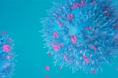
The Sanger sequencing data in our study was generated by the Sequencing and Bioanalyzer Core at BCCHR. The confocal microscopy was performed using the Leica SP8X STED confocal microscope located at BCCHR’s Imaging Core Facility. We also used the Huygens software provided by the Imaging Core Facility for image processing and deconvolution.
How did these advanced technology platforms support your research?
It was very convenient and efficient to have the Sanger sequencing done in house. Dr. Elizabeth Hui at the Sequencing and Bioanalyzer Core went above and beyond in the service she provided. Our study involved complex sequencing chromatograms and she would always offer her help to troubleshoot the chromatograms when needed.
The Imaging Core Facility at BCCHR granted us access to a state-of-the-art confocal microscope at a reasonable cost. Dr. Jingsong Wang provided extensive training for the confocal microscopy work and was always around to support our study by offering his expertise and technical assistance when required. The free image analysis workstation at the Imaging Core Facility helped us to generate high quality images suitable for publication.
Read more about this study here.
Do you have questions? Have a research story you want to share? Email core-tech@bcchr.ca.